Hi Caroline,
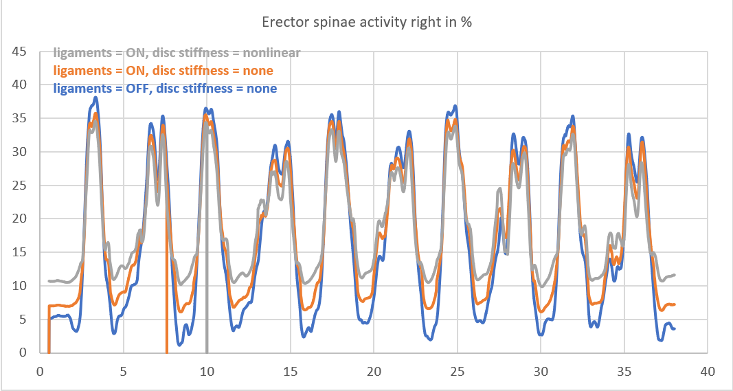
I am not sure I see the “spikes” - your curves are effectively scaled, but qualitatively are almost same. And to me it makes sense since you added some stiffness to the structure. In some case it helps muscles, in others it works in the opposite direction.
But a brief explanation: ParameterOptimization and MarkerTracking are two kinematic optimizations, where the former optimizes positions of virtual markers /sizes of individual segments to match positions/trajectories of the experimental markers, and then MarkerTracking computes angle joints curves for the whole movement based on the optimized markers and experimental data. The optimization is based on the objective function of marker error and weights parameters. Secondly, the pelvis-thorax kinematics is governed by position of the rigid thorax and pelvis thorax - effectively two rigid body rotating around each other connected by lumbar vertebrae, which are assembled into 1 kinematic constraint: the spine rhythm. The rhythm is a mechanistic way to decompose thoracopelvic angle into individual joints throughout the lumbar spine. So, what this means: you are using slightly different marker configuration - you will affect scaling of thorax, position of thorax, and this will effectively explain all the differences you see.
The ligaments and discs are effectively passive springs and they depend on computed kinematics. By optimizing movements you will get “fixed” forces/moment in these structures. These loads will, in turn, affect muscles and joint reactions, but their effect will not be “felt” by kinematics. In order, to “feel” the effect you need to include these structures into the kinematic optimization loop, which can be done by extra “soft” drivers based on potential energies of the elastic elements and changing the spine rhythm that I mentioned. To summarize: by default ligaments/discs only affect inverse dynamics loop, not kinematics.
So the question comes down to: how much do different protocols affect kinematics of the thorax? Imagine you add an extra marker exactly on top of an existing one - this will increase its weight in optimization, and the system will take into account a little more serious, because otherwise the kinematic error will increase objective function value. By adding/redistributing markers you change the sensitivity of thorax in different planes - and you will effectively get slightly different positions of thorax. But please also remember that adding markers will give a larger weight to thorax, allowing more errors in other body parts. So effectively you are looking at the sensitivity of markers right now. In our experience, we give larger weights to “reliable” markers: those that help the body part scale more reliably, and those that almost do not create any motion artifacts due to the skin sliding. The marker protocol itself is less critical than accounting for “reliable” markers.
That said, in case you had a detailed thorax, you would have more segments and having more markers would actually create a more accurate kinematic representation of the system.
And to answer your last question: try asking yourself instead how much accuracy do i need to answer my research questions? And can I explain the differences if I see them? The model has a lot of parameters and it is not realistic to find all individual stiffnesses for the ligaments/disc, muscle strengths and so on. EMG itself has similar problems - the MVC normalization may not be the ground truth, do we account for deeper muscle fibers, and so on.
I hope this helps. Kind regards,
Pavel
P.S.: I just noticed the drop in the grey plot. It is suspicious. I would closely look at this moment. Try plotting joint angles, ligament/disc loads and see what happens at this moment. You may find which object causes this behavior.